Collection:

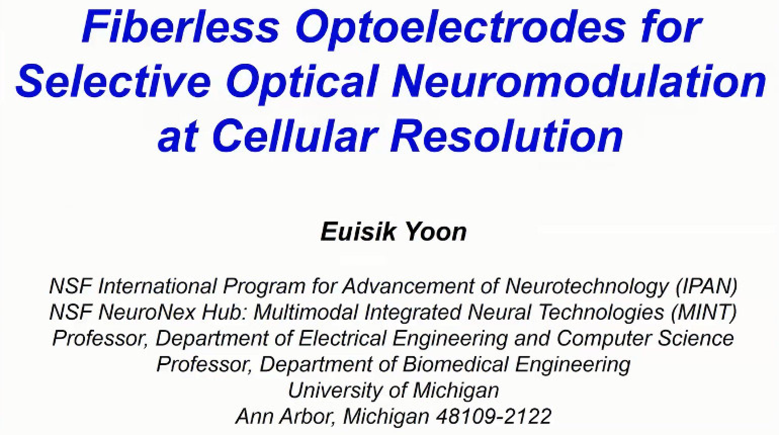
This talk will review the evolution of Michigan neural probe technologies toward scaling up the number of recording sites, enhancing the recording reliability, and introducing multi-modalities in neural interface including optogenetics. Modular system integration and compact 3D packaging approaches have been explored to realize high-density neural probe arrays for recording of more than 1,000 channels simultaneously. In order to obtain optical stimulation capability, optical waveguides were monolithically integrated on the silicon substrate to bring light to the probe shank tips. Excitation and inhibition of neural activities could be successfully validated by switching the wavelengths delivered to the distal end of the waveguide. For scaling of the number of stimulation sites, multiple micro-LEDs were directly integrated on the probe shank to achieve high spatial temporal modulation of neural circuits. Independent control of distinct cells was demonstrated ~50 ?m apart and of differential somato-dendritic compartments of single neurons in the CA1 pyramidal layer of anesthetized and freely-moving mice.
- IEEE MemberUS $10.00
- Society MemberUS $0.00
- IEEE Student MemberUS $10.00
- Non-IEEE MemberUS $20.00
Videos in this product
IEEE Brain: Fiberless Optoelectrodes for Selective Optical Neuromodulation at Cellular Resolution
This talk will review the evolution of Michigan neural probe technologies toward scaling up the number of recording sites, enhancing the recording reliability, and introducing multi-modalities in neural interface including optogenetics. Modular system integration and compact 3D packaging approaches have been explored to realize high-density neural probe arrays for recording of more than 1,000 channels simultaneously. In order to obtain optical stimulation capability, optical waveguides were monolithically integrated on the silicon substrate to bring light to the probe shank tips. Excitation and inhibition of neural activities could be successfully validated by switching the wavelengths delivered to the distal end of the waveguide. For scaling of the number of stimulation sites, multiple micro-LEDs were directly integrated on the probe shank to achieve high spatial temporal modulation of neural circuits. Independent control of distinct cells was demonstrated ~50 ?m apart and of differential somato-dendritic compartments of single neurons in the CA1 pyramidal layer of anesthetized and freely-moving mice.
 Cart
Cart Create Account
 Sign In
